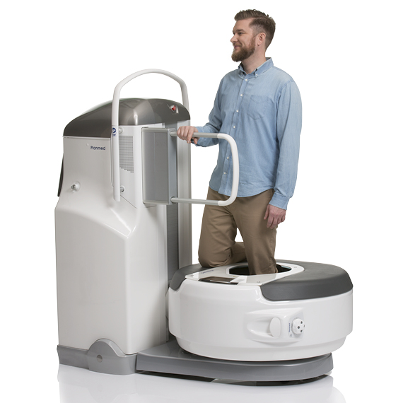
Planmed Verity
Product Description
A 3D weight-bearing image of a knee, ankle, foot, and toes under natural load can reveal problems otherwise non-discernible. The versatile patient positioning options combined with advanced imaging algorithms allow effortless imaging.
3D weight-bearing imaging solves the challenges of projection differences and overlapping structures. The anatomy is shown in a naturally occurring position and can better reveal joint space narrowing and other conditions that may not be visible through conventional diagnostic means. Extremity CT is currently the only technology able to produce 3D imaging data of the anatomy under real, weight-bearing conditions.
3D weight-bearing imaging solves the challenges of projection differences and overlapping structures. The anatomy is shown in a naturally occurring position and can better reveal joint space narrowing and other conditions that may not be visible through conventional diagnostic means. Extremity CT is currently the only technology able to produce 3D imaging data of the anatomy under real, weight-bearing conditions.
